Anatomy Of Back Of Neck Muscles - Pin On Products : Splenius capitis and cervicis proximal… muscles of back and thoracic wall.. Border of mandible and skin, and is attached to superficial fascia covering pectoralis major and deltoid muscles inferiorly. Digastric, mylohyoid, geniohyoid, stylohyoid infrahyoid muscles: There are many muscles around the neck that help to support the cervical spine and allow you to move your head in different directions. Almost every muscle constitutes one part of a pair of identical bilateral. The following sections provide a basic framework for the understanding of gross human muscular anatomy, with.
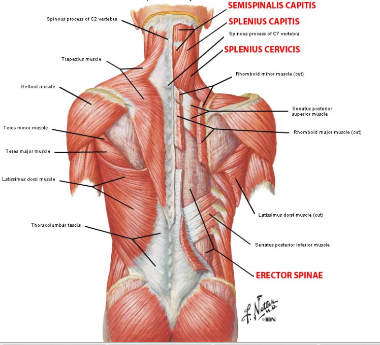
Several other muscles of the back also extend up to the neck region and are partly connected with the cervical part of the vertebral column, including the trapezius, levator scapulae, splenius, iliocostalis, longissimus, rotatores, semispinalis, interspinales, and intertransversarii muscles. Rectus capitis posterior major and rectus capitis posterior minor attach the inferior nuchal line of the occiput to the c2 and c1 vertebrae respectively. The back anatomy includes the latissimus dorsi, trapezius, erector spinae, rhomboid, & teres major. Rectus capitis, longus capitis, longus colli. Sternocleidomastoideus), with bilateral reduction throws back her head back, and at its sole tilts his head in his side (the side on which the shortened muscle).
Cervical Motor Control Part 1 Clinical Anatomy Of Cervical Spine Rayner Smale from images.squarespace-cdn.com The pll starts at c2 and goes down the back of the vertebral bodies and intervertebral discs. The splenius muscles originate at the midline and run laterally and superiorly to their insertions. Last update october 2, 2020. Digastric, mylohyoid, geniohyoid, stylohyoid infrahyoid muscles: This definition incorporates text from a public domain edition of gray's anatomy (20th u.s. Many conditions and injuries can affect the back. Watch cervical muscle anatomy animation. Border of mandible and skin, and is attached to superficial fascia covering pectoralis major and deltoid muscles inferiorly.
When we think of back muscles, latissimus dorsi (lats) comes to mind.
The muscles of the back that work together to support the spine, help keep the body upright and allow twist and bend in many directions. Splenius capitis and cervicis proximal… muscles of back and thoracic wall. It also covers some common conditions and injuries that can affect the. Muscles of the posterior neck and the back. From the sides and the back of the neck. Muscular system > muscles of neck. Human muscle system, the muscles of the human body that work the skeletal system, that are under voluntary control, and that are concerned with movement, posture, and balance. In this section, learn more about the anatomy of the muscles of the neck. Sternohyoid, sternothyroid, thyrohyoid, omohyoid anterior vertebral muscles: Equally important is the erector spinae muscles. Several other muscles of the back also extend up to the neck region and are partly connected with the cervical part of the vertebral column, including the trapezius, levator scapulae, splenius, iliocostalis, longissimus, rotatores, semispinalis, interspinales, and intertransversarii muscles. Anterior muscles of the neck. Digastric, mylohyoid, geniohyoid, stylohyoid infrahyoid muscles:
12 photos of the muscle anatomy back of neck. The suboccipital muscles act to rotate the head and extend the neck. The muscles of the back that work together to support the spine, help keep the body upright and allow twist and bend in many directions. The splenius muscles originate at the midline and run laterally and superiorly to their insertions. The back muscles stabilize and move the vertebral column, and are grouped according to the lengths and direction of the fascicles.
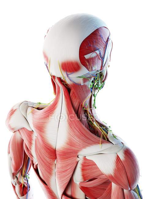
Male Back Neck And Head Muscles Computer Illustration Anatomical 3d Model Stock Photo 308626316 from st.focusedcollection.com Working in pairs on the left and. Human muscle system, the muscles of the human body that work the skeletal system, that are under voluntary control, and that are concerned with movement, posture, and balance. When we think of back muscles, latissimus dorsi (lats) comes to mind. 12 photos of the muscle anatomy back of neck. A collection of anatomy notes covering the key anatomy concepts that medical students need to learn. Bones of the neck picture. Digastric, mylohyoid, geniohyoid, stylohyoid infrahyoid muscles: Neck muscles help support the cervical spine and contribute to movements of the head, neck, upper back, and posterior longitudinal ligament (pll).
The back muscles can be three types. These types of practice studies help me to illustrate my comic art. Human muscle system, the muscles of the human body that work the skeletal system, that are under voluntary control, and that are concerned with movement, posture, and balance. Cervical spine anatomy is quite complex. The splenius muscles originate at the midline and run laterally and superiorly to their insertions. Neck muscles help support the cervical spine and contribute to movements of the head, neck, upper back, and posterior longitudinal ligament (pll). When we think of back muscles, latissimus dorsi (lats) comes to mind. A collection of anatomy notes covering the key anatomy concepts that medical students need to learn. In this section, learn more about the anatomy of the muscles of the neck. Alle muscles are detailed described incl. Chinese atlas of human anatomy. Is the only cutaneous muscle in human body (under the skin) attachments: It also covers some common conditions and injuries that can affect the.
From the sides and the back of the neck. The splenius muscles originate at the midline and run laterally and superiorly to their insertions. Together, they draw head backward, extending the neck and individually, each one draws and rotates. From the sides and the back of the neck, the splenius capitis inserts onto the head region, and the splenius cervicis extends onto the cervical region. Cervical spine anatomy is quite complex.
Building A Strong Neck T Nation from www.t-nation.com From the sides and the back of the neck, the splenius capitis inserts onto the head region, and the splenius cervicis extends onto the cervical region. Intermediate back muscles and c. The pll starts at c2 and goes down the back of the vertebral bodies and intervertebral discs. In this section, learn more about the anatomy of the muscles of the neck. The back muscles stabilize and move the vertebral column, and are grouped according to the lengths and direction of the fascicles. Muscles » muscles of the neck. Border of mandible and skin, and is attached to superficial fascia covering pectoralis major and deltoid muscles inferiorly. Intermediate layer of back muscles.
Spinous processes of txi to liii and supraspinous ligaments.
Muscles of the posterior neck and the back. Cervical spine anatomy is quite complex. Together, they draw head backward, extending the neck and individually, each one draws and rotates. This is a table of skeletal muscles of the human anatomy. There are many muscles around the neck that help to support the cervical spine and allow you to move your head in different directions. Splenius capitis and cervicis proximal… muscles of back and thoracic wall. This article covers the anatomy of the deep muscles of the back, including their function, blood supply, innervation, origin and insertion. The back has some of the body's largest muscles (erector spinae group) and some of the smallest what causes neck muscles to tighten, head stuck sideways, eyes opened, jaw locked also back here are the three classes of levers known to physics, with mechanical and anatomical examples. Last update october 2, 2020. Sternocleidomastoideus), with bilateral reduction throws back her head back, and at its sole tilts his head in his side (the side on which the shortened muscle). Intermediate back muscles and c. Rectus capitis posterior major and rectus capitis posterior minor attach the inferior nuchal line of the occiput to the c2 and c1 vertebrae respectively. There are around 650 skeletal muscles within the typical human body.
There are many muscles around the neck that help to support the cervical spine and allow you to move your head in different directions anatomy of back of neck. Together, they draw head backward, extending the neck and individually, each one draws and rotates.


0 Komentar